In This Corner
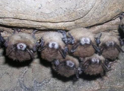
Whats Killing the Bats?
White-nose syndrome is decimating the insect-eating mammals, and researchers at the Cummings School are on the case
White Light
Optical mammography is the next frontier in breast cancer treatment
By Julia C. Keller
New technology developed at Tufts has the potential to provide diagnostic information about breast tissue in a way that could complement—and perhaps one day replace—the standard mammogram.
Sergio Fantini, a professor of biomedical engineering, uses light to obtain images of breast tissue as part of a growing field called optical mammography. This non-invasive technology could also be used repeatedly, without harm to patients, to track the effectiveness of treatment for breast cancer—the second leading cause of cancer deaths among American women.
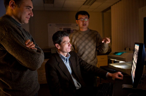
From left, Angelo Sassaroli, Sergio Fantini and Yang Yu are developing better diagnostic tools for breast cancer that could replace the traditional mammogram. Photo: Alonso Nichols
For healthy women ages 40 and older, undergoing a mammogram is a typical part of an annual physical. Having each breast compressed between two plates not once, but twice, can be an uncomfortable and even painful experience.
Early detection through x-ray mammography has saved the lives of countless women, and some men, by identifying breast cancer in its nascent stages and allowing oncologists to begin treatment earlier. Still, there are limitations to the x-ray technology. “The consensus is that x-ray mammography is very good at detecting cancer, but it’s not as good at determining, of the suspicious lesions, which ones are really cancer,” says Fantini.
If a radiologist spots something suspicious in a mammogram, a patient will then undergo a biopsy to determine whether the tissue is cancerous. If the biopsy comes back positive, the oncologist and patient can begin to make decisions about treatment.
But if the tissue isn’t cancerous, patients have gone through the emotional distress of worrying about cancer and, through the mammogram itself, have been exposed to an extra dose of tissue-damaging radiation to verify that their tissue isn’t damaged.
“Invasive biopsies or further tests like additional mammograms or having an MRI can add to the psychological stress of patients,” says Yang Yu, a biomedical engineering graduate student researcher in Fantini’s Diffuse Optical Imaging Group. “Also, you cannot take as many x-rays as you want, because it can induce cancer if you use it too much.”
Fantini’s technology may provide a solution. “Optical mammography has this huge appeal of being non-invasive and being safe,” he says.
Can’t Hold a Candle to It
The idea of passing light through living tissue to understand possible pathology can be traced back to the early 1900s. In the 1920s, the pioneers of what has become optical mammography looked for abnormal breast tissue by shining bright white light directly into the breast tissue and with the naked eye observing any problem areas.
Our eyes can perceive light wavelengths from 380 to 750 nanometers, which translate to the colors from red to violet. Near-infrared light, or NIR, falls in the 700 to 1,000 nanometer range, a slice of the light spectrum that is known as the “optical diagnostic window.” It can provide valuable information based on how tissues absorb light.
In the 1980s, researchers used NIR light to study breast tissue, which provided some improvement in discerning abnormalities. But clinicians still had difficulty in positively identifying solid breast tumors, especially when the lesions were small. It would take another decade for improvement in instrumentation and modeling to allow researchers like Fantini to explore the full range of possibilities for the technology.
Modeling how light travels through biological tissue is the expertise of Angelo Sassaroli, a research assistant professor of biomedical engineering who assists Fantini in the lab. Sassaroli describes breast tissue as a “dense cloud” that scatters photons of light in all directions. “When I inject a bunch of photons here, they travel along this cloud of trajectories. It’s not a straight line,” says Sassaroli.
By using an algorithm that Sassaroli helped Fantini refine, the researchers can create images using the intensity of the absorbed light, allowing structures like blood vessels and lesions to come into view. “With this relatively simple algorithm, we can enhance the structures within the image we see by calculating a derivative of that original image,” says Sassaroli.
Based on differences in absorption of light, NIR can be used to distinguish among water, fats and oxygen-rich and oxygen-poor tissue—the primary structures in breast tissue. “If you want to understand the interplay of these four parameters, you have to understand the fine spectral information,” says Fantini. One of the advantages of the team’s technology is the ability to obtain functional, real-time images of metabolic changes, such as blood oxygen levels, which could help pinpoint tumors.
Patience in the Exam Room
To determine if their light-based techniques can corroborate the information gathered with x-rays, Fantini is collaborating with Roger Graham, director of the Tufts Breast Center, and Marc Homer, chief of the division of mammography at Tufts Medical Center. The team recruited patients from the Tufts Breast Health Clinic to come to the lab for a proof-of-concept optical mammogram.
Their first patient is Susan, a 60-year-old woman whose x-ray mammogram revealed two suspicious lesions on her right breast. (The names of patients have been changed.) Susan comes into the lab with her daughter, Meghan, and they are met by Chia-hui Chen, E09, who is helping run the test.
Soon the scanning begins, and the software program displays real-time images as the optical system snakes back and forth, moving forward two millimeters at a time. A black-and-green graph displays the intensity of the light as data is collected. A ghostlike image of Susan’s breast begins to appear in the bottom left of the screen. Just like an exposure when developing film, the more light that passes through to the detector, the brighter the image appears. The outline of her breast is bright white, where the light has the easiest time passing through the more compressed, and therefore thinner, breast tissue. In less than five minutes, the scan is complete.
Yu, Chen and Sassaroli take another scan of Susan’s breast with the suspicious lesions, then perform two additional scans on her left breast. “These images still need to go through some processing,” says Yu. Later, Sassaroli, Fantini and Yu will process Susan’s scans using the algorithm to reveal structures like blood vessels, and hopefully the lesions, which are currently obscured.
The next steps for Fantini’s group are to work with others at Tufts, including Misha Kilmer, a professor of mathematics, and Eric Miller, an electrical and computer engineering professor and an expert in image processing and refinement. Miller and his doctoral student, Fridrik Larusson, will see if they can expand the technology’s imaging capabilities to create 3D models of the breast, rather than a two-dimensional image.
“Currently, it’s a projection—which is exactly the same thing X-ray mammography does—which isn’t ideal, because if you see blood vessels or suspicious lesions in the image, you would like to know where they are in depth,” says Fantini.
A longer version of this story appeared in the Spring 2010 issue of Alma Matters magazine.
Julia Keller can be reached at j.keller@tufts.edu.