In This Corner
Peacemakers and Rainmakers
Feinstein Center program studies the role that traditional seers play in resolving disputes in conflict-ridden eastern Africa

The Mystery of Memory
Neuroscientist Leon Reijmers is pioneering ingenious techniques to study how the brain remembers—and how it forgets
By Jacqueline Mitchell
Your first kiss, that sweatshirt you wore in college, your mother’s perfume. These can all evoke powerful memories. Like photographs, memories are the archives of our experiences. And like photographs, memories can fade over time. Despite decades of research, how the brain stores memories and discards them remains something of a scientific mystery. Leon Reijmers is working to make sense of it.
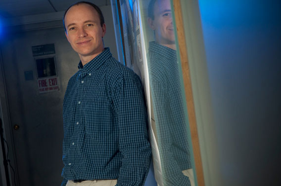
“If we can see which proteins are synthesized, we can figure out how the brain makes new memories,” says Leon Reijmers. Photo: Jodi Hilton
Reijmers, who joined the Tufts faculty this past summer as an assistant professor of neuroscience at the School of Medicine, wants to identify the specific genes and proteins involved in memory formation and discover what happens on the cellular level when we lose memories. He hopes that one day his work might lead to new therapies for those suffering from memory loss, such as Alzheimer’s patients, and those who would rather quell disturbing memories, such as victims of post-traumatic stress disorder.
Reijmers recently received a National Institutes of Health (NIH) Director’s New Innovator Award to support his research. The prestigious $1.5 million, five-year grant is intended to support creative young scientists and spur novel research with wide potential impact. Reijmers was one of only 55 chosen nationally to receive the grant.
To study how we store memories, scientists first must figure out where—among the brain’s hundred billion cells—they are stored. To do that, Reijmers and his colleagues at the Scripps Research Institute in La Jolla, Calif., where he was a post-doc, built a better mouse, which allowed them to see the specific neurons involved in memory formation.
As Reijmers reported in an August 2007 article in Science, his team created the so-called TetTag mouse by introducing a “reporter gene” into the animal’s DNA. When the mouse’s neurons fire, this gene expresses a protein that the scientists can easily detect and image. The resulting pictures provide a map of the mouse’s brain that literally highlights the neurons involved in memory formation.
Reijmers then used classical conditioning to make his TetTag mice form memories. Through mild shocks to the paws, the mice quickly learned to fear being placed in a certain box. Fear memories, says Reijmers, are well known to be stored predominately in the amygdala, an almond-shaped region of the brain associated with processing emotions.
In the 2007 proof-of-concept experiment, he was able to show that a subset of neurons in the TetTag mouse’s amygdala were indeed the ones implicated in storing this specific fear memory—they were the cells that were highlighted in the imaging when the mouse was placed in the box. Moreover, the number of neurons involved matched up to the strength of the memory: mice display fear by freezing like statues, and the more freezing behavior the TetTag mouse exhibited, the more cells were highlighted.
The project grew out of research Reijmers began as far back as college. Long interested in complex animal behavior and how the brain governs it, he began working with rats as an undergraduate in the Netherlands. Later, as a postdoctoral researcher in the lab of molecular biologist Mark Mayford at Scripps, Reijmers pioneered the use of genetic tools to pinpoint the location of memories in the brain.
Now he will build on those techniques to further investigate what goes on when the brain stores memories. The current model for memory formation suggests that the connections among neurons—called synapses—get stronger with experience; that is, the neurons become more efficient at transmitting information. But how do they do that?
Reijmers says neurons could strengthen their synapses in one or more ways. The brain cells could form more connections among each other, they could increase production of neurotransmitting chemicals or they could sprout more receptors for those neurotransmitters. Whichever process is at play here, it requires protein growth. “Over time,” says Reijmers, “if we can see which proteins are synthesized, we can figure out how the brain makes new memories. There’s still a lot we need to know about the basic mechanisms.”
The building blocks of the cell, proteins are the main ingredients in almost everything in the body. Our genes tell our cells what proteins to make, and whether those proteins become structures, like skin and hair, or chemicals, like the neurotransmitters serotonin and dopamine. Now that Reijmers’ team knows which neurons to look at, the scientists can find out which genes they express and, in turn, which proteins they synthesize during memory storage. That data will reveal whether the neurons are producing more neurotransmitters, building more receptors or creating more synapses, or some combination of the three.
The Forgotten: Suppressed or Deleted?
Another line of research focuses on what happens to neurons when a memory fades. To answer that question, Reijmers’ team—including one post-doc who started work in his lab this summer and one who will arrive this winter—will use TetTag mice that have already learned to fear the shock-box. The mice will continue to be placed in the box, but they’ll no longer receive a shock.
“Eventually, they’ll lose the fear and stop the freezing behavior,” says Reijmers. “But what is the neuronal mechanism at play? Are memories erased from the brain or just suppressed?” Images from the TetTag mouse’s brain should be instructive. If the fear-memory neurons are active, and the mouse does not freeze, that would indicate some kind of suppression. On the other hand, if the fear-memory neurons are not active, the memory is likely to have been deleted entirely from the brain.
Depending on what the data indicate, the next phase of the study will try different ways to reduce the fear behavior. Such research could be invaluable to patients with post-traumatic stress and phobias.
That’s one reason Reijmers is delighted to be at Tufts, which he calls “the perfect place to start a new lab.” As his experiments progress, he looks forward to collaborating with the university’s other neuroscience researchers as well as investigators from Tufts-affiliated hospitals.
Over the life of his New Innovator grant, Reijmer’s work will generate mountains of data, as scientists comb through each of the 20,000 to 25,000 genes in the mouse genome—which is, coincidentally, roughly the same size as the human genome. “We should find some interesting genes,” he says.
Jacqueline Mitchell can be reached at jacqueline.mitchell@tufts.edu.