In This Corner
The Creatures in the Closet
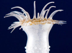
In a detective story featuring P.T. Barnum, a flood, a fire and an enterprising art historian, invaluable glasswork makes its way back home to Tufts
New Model for Biomedical Imaging
Using a different mathematical method drastically cuts computation time in determining how light travels through living tissue
By Debbie K. Chen
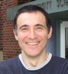
Angelo Sassaroli, a research assistant professor in the biomedical engineering department, has found a better and quicker way to model certain imaging functions.
With advances in biomedical optics—the use of light for diagnostic purposes—it’s possible to imagine that one day, for example, non-invasive biopsies could be done, eliminating the need for scalpels and recovery time. One of the major limitations in modeling how such technologies would work, though, is the computation time involved: it can take hours on a computer to model the average path of a photon of a specific wavelength as it travels through tissue.
But now Angelo Sassaroli, a research assistant professor in Tufts’ biomedical engineering department, has found a better and quicker way to model these functions, and recently published a paper on his findings in the journal Applied Optics, co-authored by colleagues at Tufts and in Italy.
Sassaroli works in Professor Sergio Fantini’s laboratory using near-infrared spectroscopy (NIRS) to study a range of topics, including brain activation, optical mammography and muscle hemodynamics, or the flow of blood through muscles. NIRS is a form of imaging that uses optical fibers to measure changes in near-infrared light as it passes through tissue, which can provide information about what lies under the skin, including cancerous activity.
NIRS is sensitive to blood flow and other parameters, but the path photons take mainly depends on the extent to which they are scattered. Even though current NIRS imaging techniques can only separate structures within tissue that are at least five millimeters apart, it can directly measure light intensity—or the amount of photons detected—due to changes inside the tissue, such as increased blood flow.
Sassaroli wanted to model the average path of photons at 250 different wavelengths, and he needed a method that would be just as accurate but faster than the Monte Carlo method, a class of computational algorithms that is the gold standard for such modeling and that is computationally intensive. To model geometries for the head and breast tissue, he used a different mathematical approach, called the Padé approximant. It turned out that in all of the models, the calculations using Padé approximants, which uses the ratio of two functions to estimate another function, were the most accurate and close to the results found using the Monte Carlo method.
The difference is that using the Padé approximants, computation time was on the order of seconds for calculating the detected light intensity at all 250 wavelengths at once. Padé’s accuracy is especially important because there is already a fast method of calculation, using perturbation theory, but it is only accurate in certain circumstances.
Sassaroli hopes that this new method will lead to better understanding of how light interacts with complex tissues. Using this knowledge, researchers hope they can improve NIRS imaging techniques and eventually be able to non-invasively and accurately evaluate the finer structures within biological tissues.
Debbie Chen is a doctoral student in the biomedical engineering department. She can be reached at debbie.chen@tufts.edu.