In This Corner
Growing Up on Camera
To create a visual history of his three children, Paul Nash photographs them every day

The Goodness of the Fit
Researcher Katya Heldwein is determined to figure out precisely how the herpes virus enters a human cell to do its damage
By Bruce Morgan
Russian-born and educated, Ekaterina Heldwein learned English during her college years from a series of educational tapes, but they featured British speakers, with proper, clipped inflections, and that was a problem. Now she was living in Portland, Oregon. When the waitress ambled over and began firing off a list of the day’s specials, none of which she had ever heard of, let alone eaten, Katya (as she is known) felt momentarily lost.
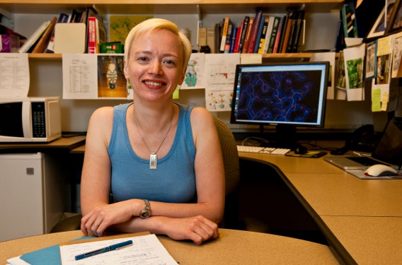
“You ask a question and get an answer, and that raises even more questions,” says Katya Heldwein. Photo: Alonso Nichols
That was OK. Her fluency lay in another direction altogether. It resided in the laboratory and classrooms at Oregon Health & Science University, where she was completing her doctorate in biochemistry and preparing to take the world of structural biology and virology by storm.
After four years as a postdoc at Harvard Medical School, working under the legendary crystallographer Stephen Harrison, and two more years as an instructor at Children’s Hospital, she landed at Tufts Medical School as an assistant professor in the Department of Molecular Biology and Microbiology in 2006 and got down to work.
Over the past four years she has walked off with just about every plum award the nation has to offer in her field. To cite just three: Heldwein was named one of 20 Pew Scholars in the Biomedical Sciences for 2007. Next she was chosen as one of 29 recipients of the New Innovator Awards by the National Institutes of Health, guaranteeing her $1.5 million in funding over five years. This spring, she received the American Society of Microbiology’s Merck Irving S. Sigal Award for young investigators in medical microbiology and infectious disease.
Her parents, still living in Russia, are surely proud. Heldwein’s mother is a textile scientist with a doctorate in chemistry. Her father has a master’s degree and an MBA. Both emphasized the importance of education to their daughter. Majoring in chemistry, Heldwein graduated from Lomonosov Moscow State University with a Red Diploma, an award for undergraduate academic excellence, in 1994.
In a way, her irregular personal trail accounts for much of her success. Heldwein’s professional starting points deviated from the norm, as her department chair at Tufts, Abraham “Linc” Sonenshein, professor of molecular biology and microbiology, explains.
“A typical background for most virologists would involve taking genetics and biochemistry and immunology—in other words, biology, biology, biology,” he says. “But Katya comes from a background in Russia, in high school and college, of having taken very high-level chemistry and physics. Her training enables her to approach biological problems in a way that most biologists are unable to do.”
Heldwein concedes the point. “My training in chemistry helps me look at the problem from a different angle,” she says, her Russian accent coloring every syllable in a way that makes you want more. Her approach involves looking at protein crystals and trying to figure out what the proteins inside look like. Her particular focus is on determining the means by which herpes viruses—human pathogens that bear a host of ills ranging from lip sores to cancer, blindness and brain inflammation—manage to enter the human cell.
A Debilitating Virus
Gary Cohen, a biologist at the University of Pennsylvania who has been collaborating with Heldwein on her research, gives some herpes background. Eight different kinds of herpes viruses can create sickness in humans, he says. Under certain conditions, they result in oral and genital herpes, the latter of which afflicts an estimated 30 percent of the U.S. population. Herpes ranks as the leading cause of viral blindness and viral encephalitis in the country.
But there’s much more. The cytomegalovirus promotes organ transplant rejection. The varicella-zoster virus yields childhood chicken pox and its adult variant, shingles, a painful and debilitating nerve ailment. The herpes family also includes the Epstein-Barr virus, which causes Burkitt’s lymphoma, and Kaposi’s sarcoma, a skin cancer common in AIDS patients. What we have here are troubling, even lethal afflictions, all bundled into one nasty package, poised and waiting to do harm.
“If the virus cannot get in, there’s no infection,” Cohen notes. “Once the precise entry mechanism is determined, then we have a target and a strategy for heading off the threat.” Rebuffed at the membrane, the protective barrier of the cell, the herpes virus would become one of many viruses circulating harmlessly throughout the body, like a distracted burglar who isn’t able to crack a building’s security and so wanders endlessly around its perimeter.
The process by which a herpes virus gains entry into the human cell would seem an obvious topic of interest to virologists, but when Heldwein began work at Harvard, she recounts, “I was floored that people who have studied this for many years didn’t have answers to even basic questions.” For starters, what do the four glycoproteins that collaborate to let the virus into the cell look like? And, more importantly, what is it that they do, exactly, and how do they do it? Perhaps surprisingly, the key to the first question helps unlock the next two.
Structural biology explores the relationship between form and function at the atomic level. It’s supremely logical in its approach. Since no biological detail in nature is wasted, everything you see has a purpose. Think about it this way: If you came across a hammer for the first time, it wouldn’t take long to figure out what it does. Similarly, by first determining the structure of molecules, scientists like Heldwein can work backwards, in effect, to deduce their function. As she puts it, with elegant simplicity, “What something does depends on what it looks like.”
Consider a race car and a tractor side by side, she suggests. “If you look at the race car and the tractor, you can tell that they’re designed to do different things.” The same is true of a protein’s shape and form. Some functions you can rule out, based on your knowledge or experience, and then, by process of elimination, you can further narrow the field of possibilities. “Once you have the structure of a protein, you can say, ‘Well, maybe our protein is capable of doing this.’ Then you do experiments in the lab to find out,” Heldwein says.
For whatever reason, Heldwein has this rarefied field of race cars, tractors and protein atoms largely to herself. In the first place, structural biology occupies a relatively small corner within the field of biology. Then the number of specialists in virology has never been big. The number of structural biologists who have also trained in virology can be counted on one hand, says Heldwein, before considering a moment. “Well, maybe two hands,” she laughs. She is, by every pertinent measure, including her birthplace, her training and her professional focus, an oddity.
Navigating the Atomic Neighborhood
Let’s presume we want to know what the four glycoproteins that let the herpes virus into the human cell look like, so that we can figure out their function and, with any luck, their means of attack. What’s so hard about getting a look at a few proteins? Roselyn Eisenberg, a professor of microbiology at the University of Pennsylvania and the second of Heldwein’s two chief collaborators there, outlines the problem.
“If you put a protein under a conventional microscope, you’ll see almost nothing,” she says. “They are that small in size. It’s like if you take a picture of some kids from atop a nearby building—you’ll see that they’re there, but without any detail. If you want to see them in focus, you need other tools. Structural biologists always want to bring proteins into focus so they can see what they look like and see how they work.”
A standard lab microscope can magnify something a thousand times, Eisenberg says. Even a much fancier electron microscope has a limit of about 500,000 times magnification—“still not big enough.” That leaves the synchrotron, a high-energy particle accelerator that provides the means to obtain the best view possible of an individual protein. There are perhaps half a dozen of these devices in the U.S., and they are in high demand. Heldwein uses one at the Argonne National Laboratory, near Chicago.
The synchrotron can yield X-ray diffraction images of individual protein crystals, but they are not actual photographs. Normally, when light passes through a lens, it is focused by the lens material. In a camera, this would be a wafer of precision-ground glass. But that is not possible here. “There is no focusing lens, because there is no material that can do it,” says Heldwein. What this means is that the synchrotron does not produce a coherent picture at the receiving end. Instead, as the X-rays pass through the crystal, they encounter electrons and scatter in different directions, projecting an intricate pattern of dots onto a receiving “image plate.”
“We can’t exactly see the structure of the protein, but we get these X-ray diffraction images,” Heldwein says. “It’s kind of like a photograph, but not a true representation.” In the words of Gary Cohen, who has been explaining the steps in the process, “If you’re lucky and you get the crystal to diffract, then you’ve got a series of hoops you have to jump through.”
Advanced math must do the work of the missing camera lens. Heldwein proffers a page of typical equations used to do the job. A quick warning for the math-averse: This is not the kind of math you remember from school, where two trains are speeding toward each other on parallel tracks, or a triangle needs to have the length of its sides computed. This is instead the kind that you might find crowding the blackboard if you pushed open a door in a doctoral class in physics at MIT.
The Art of Crystals
Heldwein explains that each spot in the X-ray diffraction image contains information about all the electrons within the crystal, much as any cell in the body holds data for all the rest. There are thousands of spots resulting from each diffraction experiment, and each spot in turn can be plotted for three factors: location, density and phase. Heldwein is quick to point out that the actual result from a series of diffraction experiments and subsequent calculations is not the protein structure, per se, but rather a 3-D picture of electron distribution.
Electrons circling the atoms are elusive, and, in accord with the Heisenberg Uncertainty Principle, which states that an electron’s location and momentum cannot be determined simultaneously, impossible to fix in place. A complex software program is used to display the electrons’ likely location, something like their approximate path through the atomic neighborhood.
Electron density maps that result from these calculations appear as tubular clouds. The next challenge is to find locations for all the atoms in the protein in relation to one another on this map. How well the locations of atoms match the electron path is known, in a curious phrase that sounds like a bad translation, as “the goodness of the fit.”
For all of this deductive and reductive magic to work, a potent blend of technical prowess and brainpower is needed, says Eisenberg.
“There is an art to making crystals, and then they must diffract well, and they must survive in the beam,” she says. “Katya’s very skilled at this kind of bench work. The process takes patience, because until you get to the end, meaning the electron density map, you have nothing. When you finally reach the point of building a model of the protein structure, you need to use everything you know about amino acids and how they behave. You have to think in three dimensions. Interpreting the data takes a lot of creativity, a lot of intelligence—and that’s the real skill, the interpretation.”
So far, Heldwein and her collaborators have determined the structures of all four herpes virus proteins. “Now we need to understand how they work together,” says Heldwein. “Why are there four? We don’t know. We can speculate, but we don’t know.” She laughs. “The viruses know better than we do.” She offers this last remark with a humble, respectful edge, as she always does when talking about human viruses. “To be honest, they’re smarter than we are,” she says another time, her eyes widening at the fact of it.
Maybe so, but Heldwein is closing in on her goal. For example, one of the four proteins in the herpes virus cluster shows an unusual structure. “It doesn’t look like anything else out there,” the virologist notes. “So what does it do?” In its outline, the protein resembles a boot. Preliminary tests suggest that its function may be to activate a second protein into doing its job of cell entry.
“It acts like the ‘mover and shaker’ of this other protein,” she says. “It looks like a boot and sort of ‘kicks’ the fusion protein into action.” Heldwein savors the apparent link between the protein’s appearance and function, calling it “very satisfying, aesthetically.”
What shines through when you spend time in Heldwein’s company is her way of thinking concretely about the structure and function of proteins—the same way that you and I might think about traffic cones on a highway, or cartons on a shelf—and her absolute immersion in the mysteries there. She gets pumped thinking and talking about the subject. It’s mostly a pent-up, internal combustion sort of thing going on in her, but she can spread the heat, too. An otherwise bone-dry report from a scientific conference held at Cornell in 2004 confirmed the reach of her infectious personal style by noting the “captivating presentation” she had given.
Heldwein, Cohen and Eisenberg collectively have reached a thrilling moment in their professional lives. Never before have the four proteins that make up the herpes virus been visible the way they are right now. Cohen, for one, can’t contain his enthusiasm. “Oh, it’s unraveling,” he says excitedly. “It’s like you’ve got a puzzle with a picture on the cover of the box. As soon as you see what the pieces look like, you can start fitting in a piece. You cram one piece in—it fits, but not exactly. So you still don’t know what’s going on, but piece by piece you can get there.”
When Cohen’s zeal is relayed to her, Heldwein laughs. “That’s Gary,” she says. “Is the work getting easier or harder now? I think it’s getting more complex. But that’s true everywhere in science. You ask a question and get an answer, and that raises even more questions.”
This story first appeared in the summer 2010 Tufts Medicine magazine.
Bruce Morgan, can be reached at bruce.morgan@tufts.edu.